Canine Karyotype
Many human cancers are associated with characteristic chromosome aberrations that have diagnostic and prognostic significance. The study of chromosomes (cytogenetics) and the identification of chromosome aberrations now plays a key role in defining many human cancers and in selecting appropriate therapy. The knowledge of such aberrations has identified areas of the human genome to be targeted for further research. In the dog the extent and identity of chromosome aberrations associated with specific cancers is still largely unknown. Historically, this has been a consequence of the difficulties in identifying reliably the chromosome comprising the dog karyotype. However, with an internationally recognized chromosome nomenclature for the dog, researchers now have the ability to maintain consistency in canine cytogenetic descriptions. A more recent tool for the classification of many human tumors has been provided by cellular genomics, in particular by molecular cytogenetic evaluation using fluorescence in situ hybridization (FISH) techniques. In the human, many cancers are actually now defined by their recurrent genetic abnormalities. These include; whole chromosome numerical abnormalities (e.g. trisomies and monosomies); partial chromosome numerical abnormalities (e.g. duplications and deletions); and structural abnormalities (e.g. translocations). Molecular cytogenetic evaluation of tumors using FISH has revolutionized the way in which we interrogate the cancer genome for recurrent changes. The FISH techniques widely used in cancer studies include metaphase FISH, interphase FISH and comparative genomic hybridization (CGH). All three techniques play an important role in the molecular cytogenetic evaluation of tumors.

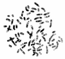
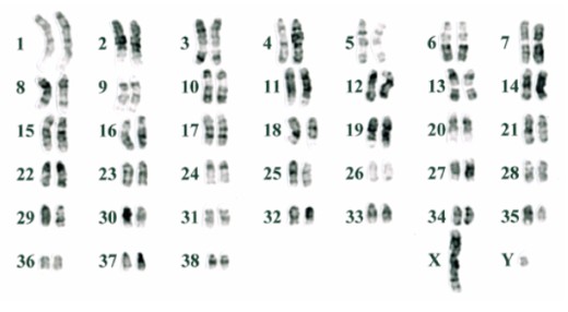
Using multicolor FISH we are able to conclusively identify each chromosome pair and generate a karyotype of the above cell, as shown below.
